摘要
目的:
1.探究超声引导下细针穿刺细胞学检查(ultrasound-guided fineneedle aspiration cytology, US-FNAC )与常规超声(Coventionalultrasound, CUS)评估甲状腺结节的临床价值,以及比较 US-FNAC对不同直径大小结节的诊断价值。
2.探讨 US-FNAC 在甲状腺影像报告和数据系统(Thyroidimaging reporting and data system,TI-RADS)3、4a 类结节良恶性鉴别诊断中的应用价值,进一步改善甲状腺结节的临床 TI-RADS 分级。
3.分析甲状腺结节患者的临床及病理学特点,探讨甲状腺恶性结节的相关危险因素,辅助诊断甲状腺结节的性质。
方法:
回顾性分析 2018 年 1 月--2019 年 1 月收治于南华大学附属第一医院乳甲外科 121 例甲状腺结节患者的病例资料,所有患者均行CUS、US-FNAC 检查、血清甲状腺功能检测以及手术治疗,结节性质以手术后组织病理学结果为金标准,分别进行以下对比:
1.比较 CUS 和 US-FNAC 检查单独诊断甲状腺结节良恶性的灵敏度、特异度、诊断准确度、假阳性率、假阴性率,进一步比较US-FNAC 对甲状腺结节直径≤10mm 组和>10mm 组的诊断效能。
2.采用χ2检验计算 FNA 细胞学诊断 TI-RADS 3、4a 类结节的灵敏度、特异度、准确度、阳性预测值、阴性预测值,以 P<0.05 为差异有统计学意义。
3.根据术后组织病理结果将其分为两组,良性组(良性结节)35例,恶性组(恶性结节)86 例。单变量分析两组患者的年龄、性别、血清甲状腺球蛋白抗体(Thyroglobulin antibody,TgAb)、促甲状腺激素(Thyroid stimulating hormone,TSH)、游离甲状腺素(FreeThyroxineIndex,FT4 ) 、 游 离 三 碘 甲 状 腺 原 氨 酸 ( Freetriiodothyronine,FT3)水平、FT4/FT3 的比值等结果;然后将统计学上有意义的单因素指标纳入多变量 logistic 回归分析,筛选出恶性甲状腺结节的危险因素。
结果:
1.US-FNAC 诊断阳性结节 100 例(恶性 79 例+可疑恶性 21 例);阴性结节 21 例(良性 18 例+意义不明确的细胞非典型病变(atypia ofundetermined significance,AUS)3 例);其准确度、灵敏度、特异度、假阳性率、假阴性率分别为:85.9%(103/121)、97.7%(84/86)、54.3%(19/35)、45.7%(16/35)、2.3%(2/86)。超声评估诊断阳性结节 101 例,阴性结节为 20 例;其准确度、灵敏度、特异度、假阳性率、假阴性率分别为 69.4%(84/121)、87.2%(75/86)、25.7%(9/35)、74.3%(26/35)、12.8%(11/86);US-FNAC 的准确度和灵敏度均高于普通超声,假阴性率相对较低,差异有统计学意义(P<0.05)。≤10mm 结节和>10mm 结节两组间 US-FNAC 的特异度、阴性预测值和诊断符合度差异有统计学意义(P<0.05)。
2.本研究有 TI-RADS 3 类结节 22 例,FNA 细胞学诊断结果为阳性结节 12 例,阴性结节 10 例;TI-RADS 4a 类结 84 例,其 FNA 细胞学诊断结果为阳性结节 73 例,阴性结节 11 例;与术后病理结果(3类:恶性 12 例+良性 10 例;4a 类:恶性 61 例+良性 23 例)对比,差异均有统计学意义(P<0.05)。US-FNAC 诊断的灵敏度、特异度、准确度及实际恶性率分别为 TI-RADS 3 类结节 83.3%、80.0%、81.8%、54.5%;TI-RADS 4a 类结节:灵敏度、特异度、准确度及实际恶性率分别为 78.1%、90.9%、79.8%、72.6%。在诊断恶性结节时,US-FNAC 对 TI-RADS 3 类结节的诊断评估比 TI-RADS 4a 类结节更准确和敏感。
3.单变量分析结果显示,良恶性组的年龄、TgAb、TSH 和FT4/FT3 的比值有统计学差异(P<0.05)。多变量 logistic 回归分析结果显示年龄和 TgAb 水平是甲状腺恶性结节的独立危险因素(P<0.05)。
结论:
1.US-FNAC 鉴定甲状腺结节性质的准确度高于常规超声。
2.甲状腺结节患者的年龄、血清 TSH、TgAb、FT4/FT3 比值可能对鉴别甲状腺结节性质具有较高的辅助诊断价值。
3.US-FNAC 对于 TI-RADS 3、4a 类结节的良恶性测定具有较高的诊断效能,有利于减少对 TI-RADS 3、4a 类结节的低估。
关键词: 细针穿刺;超声检查;甲状腺结节;甲状腺影像报告与数据系统。
Abstract
Objective:
1. To explore the clinical value of ultrasound-guided fine needle aspiration cytology and conventional ultrasound evaluation of thyroid nodules,and to compare the diagnostic value of US-FNAC for nodules of different diameters.
2. To investigate the fine needle aspiration (FNA) in the thyroid imaging reporting and data system (thyroid imaging reporting and Data system, TI-RADS) The value of the differential diagnosis of benign and malignant nodules of type 3 and 4, further improve the clinical TI-RADS classification of thyroid nodules.
3. To analyze the clinical and pathological features of patients with thyroid nodules, to explore the risk factors associated with malignant thyroid nodules, and to assist in the diagnosis of thyroid nodules.
Methods:
Retrospective analysis of 121 cases of thyroid nodules in the Department of Breast Surgery, the First Affiliated Hospital of University of South China from January 2018 to January 2019. All patients underwent CUS, US-FNAC, serum thyroid function tests, and Surgical treatment, the nature of the nodules was based on the post-operative histopathological results as the gold standard, and the following comparisons were made:
1. Compare the sensitivity, specificity, diagnostic accuracy, false positive rate, and false negative rate of the diagnosis of benign and malignant thyroid nodules by CUS and US-FNAC respectively.Further compare US-FNAC to thyroid nodules ≤10mm and >10mm Diagnostic efficacy.
2. The sensitivity, specificity, accuracy, positive predictive value and negative predictive value of FNA cytological diagnosis of TI-RADS type 3 and 4a nodules were calculated by χ 2 test. The difference was statistically significant at P<0.05.
3. According to the postoperative histopathological results, they were divided into two groups: benign group (benign nodules) in 35 cases, malignant group (malignant nodules) in 86 cases. Univariate analysis of age, sex, serum thyroglobulin antibody (TgAb), thyrotropin (TSH), free thyroxine (FT4), free triiodothyronine (FT3) levels, the FT4/FT3 ratio,which the results were compared, and then multivariate logistic regression analysis was used to screen the risk factors of malignant thyroid nodules.
Results:
1. US-FNAC diagnosed 100 cases of positive nodules (79 cases of malignant nodules + 21 cases of suspected malignancy); 21 cases of negative nodules (18 cases of benign nodules + atypia of undetermined significance (AUS) 3 cases); its accuracy, sensitivity, specificity, false positive rate, false negative rate are: 85.9% (103/121), 97.7% (84/86),54.3% (19/35), 45.7% (16/35), 2.3% (2/86). Ultrasound was evaluated as positive nodules in 101 cases and negative nodules in 20 cases. The accuracy, sensitivity, specificity, false positive rate and false negative ratewere 69.4% (84/121) and 87.2% (75/86), respectively. 25.7% (9/35),74.3% (26/35), 12.8% (11/86); The accuracy and sensitivity of US-FNAC were higher than those of ordinary ultrasound, and the false negative rate was relatively low, and the difference was statistically significant (P<0.05).The specificity, negative predictive value and diagnostic compliance of US-FNAC between the nodules ≤10mm and >10mm were statistically significant (P<0.05).
2. In this study, there were 22 cases of TI-RADS type 3 nodules. The results of FNA cytology diagnosis were positive nodules in 12 cases,negative nodules in 10 cases, and TI-RADS 4a type in 84 cases. The FNA cytological diagnosis results in positive nodules. 73 cases, 11 cases of negative nodules; compared with postoperative pathological results (3 categories: malignant 12 cases + benign 10 cases; 4a: malignant 61 cases + benign 23 cases), the difference was statistically significant (P <0.0. 05).The sensitivity, specificity, accuracy, and actual malignancy of US-FNAC diagnosis were 83.3%, 80.0%, 81.8%, and 54.5% of TI-RADS type 3 nodules, respectively; TI-RADS type 4a nodules: sensitivity,specificity, Accuracy and actual malignancy were 78.1%, 90.9%, 79.8%,and 72.6%, respectively. In the diagnosis of malignant nodules,US-FNAC's diagnostic assessment of TI-RADS Class 3 nodules is more accurate and sensitive than TI-RADS Type 4a nodules.
3. Univariate analysis showed that the age, TgAb, TSH and FT4/FT3 ratios of the benign and malignant groups were statistically different (P<0.05). Multivariate logistic regression analysis showed that age and TgAb levels were independent risk factors for malignant thyroid nodules (P<0.05).
Conclusion:
1. US-FNAC identifies the nature of thyroid nodules with higher accuracy than conventional ultrasound.
2. The age, serum TSH, TgAb, FT4/FT3 ratio of patients with thyroid nodules may have a higher diagnostic value for the identification of thyroid nodules.
3. US-FNAC has a high diagnostic efficiency for the benign and malignant determination of TI-RADS 3, 4a nodules, which is beneficial to reduce the underestimation of TI-RADS 3, 4a nodules.
Key Words: Fine needle aspiration ultrasonography thyroid nodules thyroid imaging report and data system。
第1章 前 言
甲状腺结节是甲状腺常见病和多发病,近年来,随着人们生活饮食习惯和周边环境的改变,甲状腺结节的发病率也反应性地呈现出一种攀升甚至年轻化的趋势,对人们的生命安全构成严重威胁[1]。大多数甲状腺结节是良性结节,约占颈部肿块的 50%[2],而恶性甲状腺结节的发现率仅为 5%-10%。甲状腺癌主要分为 4 类:甲状腺乳头状癌(papillary thyroid carcinoma,PTC)、甲状腺滤泡状癌(follicular thyroid carcinoma,FTC)、甲状腺髓样癌(medullarythyroid carcinoma,MTC)和甲状腺未分化癌(anaplastic thyroid cancer,ATC);其中,临床上以 PTC 较为常见,约占甲状腺癌的 80%-85%,其预后相对较好。因此,确定甲状腺结节性质是临床诊断和治疗的重点。
诊断甲状腺疾病有许多医学检查手段,临床上可能先是门诊医师通过触诊的方式进行判断,但这种方式的主观性太大,并且部分小结节易被忽略,容易造成漏诊。此外,我们还可以使用甲状腺激素检测、甲状腺 B 超、计算机断层扫描或核磁共振、超声引导下甲状腺细针穿刺细胞学检查(ultrasound-guided fine needle aspiration cytology,US-FNAC)等相关检查来发现甲状腺结节;高分辨率超声是目前评估甲状腺结节的首选方法,但由于甲状腺结节错综复杂,良性和恶性结节的超声图像缺乏特异性,且超声医师的工作经验和主观性很大程度地影响了检查结果,故而,超声检查具有一定的局限性。
甲状腺穿刺活检是临床上不可或缺的检查方法,起初,甲状腺穿刺活检可分为粗针穿刺和细针穿刺两种方法, 粗针穿刺的优点是获得腺体组织多, 但对正常甲状腺组织损伤较大, 穿刺出血的风险高, 而且甲状腺结节一般较小,它的穿刺阳性率低,遂逐渐淡出临床诊疗工作。20 世纪 80 年代初,甲状腺细针穿刺就被应用于欧洲及北美一些医学中心[3, 4],它又可分为超声引导下甲状腺细针穿刺细胞学检查(US-FNAC)和触诊下甲状腺细针穿刺活检(Freehand fine needle aspiration biopsy,Freehand-FNAB),但US-FNAC 具有创伤小、耗时短、操作简单安全等优势,已然成为检查甲状腺结节性质不可或缺的手段。在国内有相关研究称,US-FNAC 对恶性甲状腺结节的灵敏性和特异性超过 95%以上[5]。尽管甲状腺穿刺细胞活检术有诸多优势,但它也受制于操作者的熟练程度以及结节大小、涂片技术等多因素影响,所以有一定的局限性。临床研究表明,TI-RADS 3 类结节的恶性风险指数小于 5%,而 TI-RADS 4 类结节的恶性风险指数为 5%-85%,其中4a 类结节的恶变风险为 5%-10%,两者恶变风险差异较小,在临床上可能出现漏诊的情况,因此结合术前血清甲状腺功能检测也是一项必不可少的方法。促甲状腺激素(Thyroid stimulating hormone,TSH)是促进甲状腺细胞生长和调节甲状腺功能的因子,近年来,研究发现甲状腺癌患者的 TSH 水平高于良性甲状腺结节患者[6, 7],但也有学者对此存在争议[8, 9]。此外,还有学者认为甲状腺球蛋白抗体(Thyroglobulin antibody,TgAb)和游离甲状腺素(Free ThyroxineIndex,FT4)可作为预测甲状腺癌的诊断指标[10, 11]。另外关于用 FT4/FT3 比值鉴别结节良恶性的研究鲜有报道。
总而言之,超声引导下甲状腺细针穿刺细胞活检术对于鉴别结节性质和随后的治疗具有重要的临床价值。本课题旨在进一步研究和探讨超声引导下甲状腺细针穿刺细胞活检术与血清学检查对鉴别甲状腺结节性质的临床应用价值。
第2章 资料及方法。
2.1 病例资料。
收集 2018 年 1 月--2019 年 1 月南华大学附属第一医院乳甲外科 121 例甲状腺结节手术患者的病例资料,包括术前超声检查结果、穿刺细胞学结果、术前甲状腺功能以及术后病理结果。
2.2 病例纳入标准。
2.2.1 入选标准。
(1)符合甲状腺结节细针穿刺的活检指征(《2016 年版美国甲状腺协会(ATA)指南》[12]、《超声引导下甲状腺结节细针穿刺活检专家共识及操作指南(2018 年版)》[13]);(2)术前均行 US、血清甲状腺功能检测、US-FNAC 检查,并有术后组织病理学证实者;(3)甲状腺从未接受过微波消融、I131 放射、手术等相关治疗;(4)既往无头颈部恶性肿瘤放疗病史;(5)无肝、肺等远处转移征象;(6)门诊及住院病例资料完整。
2.2.2 排除标准。
(1)存在穿刺检查或甲状腺切除术禁忌症;(2)既往有甲状腺手术史;(3)具有出血倾向,出、凝血时间明显延长,凝血酶原活性显着减低;(4)严重高血压、糖尿病或重要器官功能不全等内科慢性疾病者;(5)合并其他恶性肿瘤者;(6)合并精神疾病或障碍者。
2.3 仪器和设备。
(1)诊断仪器为东芝 Aplio500 超声诊断仪,探头为浅表探头 PLT-805AT,频率设定为 10MHz;(2)穿刺仪器为 Mindray UMT-150 超声诊断仪,探头为浅表探头 7L4P,频率设定为 10MHz,并配备穿刺引导装置;(3)23G 一次性穿刺针。
2.4 检查方法。
2.4.1 CUS 检查方法。
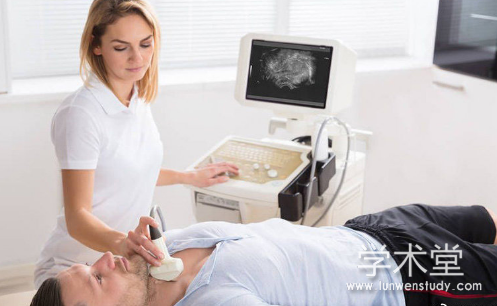
将患者置于仰卧位, 充分暴露颈前部区域, 于肩膀和颈下垫枕, 颈部保持斜躺状态。由专业超声科医师对患者左、右两个侧叶、峡部及双侧颈部淋巴结进行常规超声检查并存储结节病灶图像, 根据甲状腺影像和数据系统(Thyroidimaging reporting and data system,TI-RADS)对甲状腺结节进行评估分级。
2.4.2 US-FNAC 检查方法。
(1)穿刺前准备:核对患者信息和结节信息、询问患者的病史和既往病史、评估一般健康状况、解释穿刺操作风险和注意事项、并签署有创操作同意书。
(2)穿刺取材:嘱患者取仰卧位,颈肩部垫高呈过伸位;颈部常规消毒,铺无菌洞巾,超声探头无菌处理后定位甲状腺可疑结节,在超声探头监视下采用非负压吸引穿刺方法,以穿刺点为中心快速经皮进针穿刺进入甲状腺结节,并保持穿刺针长轴与声束在同一平面,注意避开较明显的血管,在可疑结节内从不同部位、不同方向反复提插和抽吸 3~5 次,立即拔出穿刺针完成取材。
(3)标本立即固定、涂片:将提取的组织样本快速涂片(4-6 张),用 95%的乙醇固定,并打包送往病理科行细胞学检查。注意观察标本是否符合细胞学诊断要求,若吸取组织较少,可对该结节再一次行穿刺。
(4)穿刺完毕后,对穿刺点予以消毒并覆盖敷贴,在观察区留观 20~30 min,适当按压穿刺点止血 20~30 min,并向病人交代穿刺后注意事项。
2.4.3 甲状腺功能检测方法。
术前采集甲状腺结节患者清晨空腹外周静脉血,检测外周血中 FT3、FT4、TSH、TgAb 水平,并计算 FT4/FT3 的比值。
【由于本篇文章为硕士论文,如需全文请点击底部下载全文链接】
第 2 章 资料与方法
第 3 章 结果
第 4 章 讨论
附图
第5章 结 论
1.US-FNAC 鉴定甲状腺结节性质的准确度高于常规超声。
2.甲状腺结节患者的年龄、血清 TSH、TgAb、FT4/FT3 比值可能对鉴别甲状腺结节性质具有较高的辅助诊断价值。
3.US-FNAC 对于 TI-RADS 3、4a 类结节的良恶性测定具有较高的诊断效能,有利于减少对 TI-RADS 3、4a 类结节的低估。
参考文献