图4(b)为SV算法得到的牛奶流强度图像,加入多普勒频移算法得到的牛奶流方向信息可以得到含有牛奶流强度和方向的图像,即图4(d)。图4(d)中不同的颜色表示牛奶分子不同的运动方向,证明了综合应用SV算法与多普勒OCT算法确实能够获得具有方向信息的流体强度图。图4(d)与图4(c)相比,大部分背景噪声被消除,证明提出的方法确实能够消除背景噪声,改善成像质量。利用SV算法获取良好强度信息的优势和多普勒OCT算法获取速度方向的优势,既很好地消除了背景噪声,又获得了方向信息。后续对所有帧(B扫)进行同样处理,经过整合可以得到三维成像结果。
3.2人体皮肤活体实验。
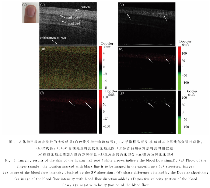
在实验中,对人体左手无名指上靠近指甲根部的皮肤处用SSOCT进行成像,每个B扫(x方向)中包含1000个A扫,每个C扫(y方向)中包含400个B扫,选取其中效果较好的B扫图像进行算法处理,结果如图5所示。
图5(a)黑线标示的位置为手指的指甲根部部分,图5(b)为SSOCT系统得到的指甲根部皮肤处的结构图以及实验中加入的另一片平面参考镜的像。通过SV算法处理得到血流的强度图为图5(c)。在显示经校正的多普勒频移图像时,通过伪彩色增强映射至-6~6区间,其中0定义为黑色,代表静止的无多普勒频移的位置;定义-6~0为负向多普勒频移,从绿色渐变为黑色,数值越小,代表的频移越大;定义0~6为正向多普勒频移,从黑色渐变为红色,数值越大,代表的频移越大。利用多普勒OCT算法得到的图像即图5(d)。将图5(d)中得到的多普勒频移Δf信息引入血流强度图5(c)中得到具有方向信息的血流强度图像,即图5(e),通过伪彩色映射到-150~150区间,-150~0表示负方向,0~150表示正方向。图5(f)和图5(g)表示多普勒频移算法得到的血流正负流向的信息,其伪彩色映射区间同图5(e)。
SV算法处理结构图得到血流强度图像,即图5(c),成功地除去了固体组织的部分,提取出了血流信号。多普勒OCT算法得到的图像由于背景噪声的影响,较难进行辨识,但提供了血流的方向信息。对图5(c)加入改进的多普勒频移算法得到的血流方向信息后得到了具有方向信息的血流强度图像,即图5(e)。图5(e)不同颜色的斑点表示不同流向的血管截面图。图5(f)和图5(g)分别显示了正向和负向的血管截面图。后续对所有帧(B扫)进行同样处理,可以得到三维血流成像结果。
4结论。
基于手持式SSOCT系统提出一种提取血流信号强度和方向的方法,即综合应用SV算法和多普勒算法,对于其中多普勒算法部分中时间引入的相位跃变,实验系统中加入了一面标准平面镜(在与样品干涉范围之外)用来消除由于扫频光源触发和采集卡采集之间时间延迟变化带来的相位跃变。在不消减两种方法各自优势的前提下,得到一幅同时含有血流信号强度和方向的图像,经过多帧重复计算处理,最终可以得到人体皮肤三维血流强度和方向信息的图像。结果表明,部分噪声会对血流信号的分辨产生干扰,有待后续的改进。
参考文献:
[1]Huang D,Swanson E A,Lin C P,et al.Optical coherence tomography[J].Science,1991,254(5035):1178-1181.
[2]Zhu Hailong.Swept source optical coherence tomography system based on LabVIEW[D].Hangzhou:Hangzhou DianziUniversity,2013.朱海龙。基于LabVIEW的扫频OCT系统研究[D].杭州:杭州电子科技大学,2013.
[3]Shi Weisong,Gao Wanrong,Chen Chaoliang.Handheld swept source optical coherence tomography for imaging humanskin in vivo[J].Acta Optica Sinica,2015,35(11):1117001.史伟松,高万荣,陈朝良。人体皮肤在体手持式扫频光学相干层析系统[J].光学学报,2015,35(11):1117001.
[4]Wu Tong,Ding Zhihua.Development of 20kHz swept source optical coherence tomography system[J].Chinese JLasers,2009,36(2):503-508.吴彤,丁志华。20kHz扫频光学相干层析系统[J].中国激光,2009,36(2):503-508.
[5]Wang Ling,Zhang Lielie,Zhou Qingqing,et al.OCT-based improvement of geometrical controllability of 3D-bioprinted porous hydrogel scaffolds[J].Chinese J Lasers,2016,43(6):0607001.王玲,张烈烈,周青青,等。基于光学相干层析的水凝胶三维打印精准控制研究[J].中国激光,2016,43(6):0607001.
[6]He Qiyu,Li Zhongliang,Wang Xiangzhao,et al.Automated retinal layer segmentation based on optical coherencetomographic images[J].Acta Optica Sinica,2016,36(10):1011003.贺琪欲,李中梁,王向朝,等。基于光学相干层析成像的视网膜图像自动分层方法[J].光学学报,2016,36(10):1011003.
[7]Blatter C,Weingast J,Alex A,et al.In situ structural and microangiographic assessment of human skin lesions withhigh-speed OCT[J].Biomedical Optics Express,2012,3(10):2636-2646.
[8]Mariampillai A,Leung M K K,Jarvi M,et al.Optimized speckle variance OCT imaging of microvasculature[J].Optics Letters,2010,35(8):1257-1259.
[9]Zhang A Q,Zhang Q Q,Chen C L,et al.Methods and algorithms for optical coherence tomography-basedangiography:a review and comparison[J].Journal of Biomedical Optics,2015,20(10):100901.
[10]Wang R K,Jacques S L,Ma Z H,et al.Three dimensional optical angiography[J].Optics Express,2007,15(7):4083-4097.
[11]Barton J K,Stromski S.Flow measurement without phase information in optical coherence tomography images[J].Optics Express,2005,13(14):5234-5239.
[12]Mariampillai A,Standish B A,Moriyama E H,et al.Speckle variance detection of microvasculature using swept-sourceoptical coherence tomography[J].Optics Letters,2008,33(13):1530-1532.
[13]Jonathan E,Enfield J,Leahy M J.Correlation mapping method for generating microcirculation morphology from opticalcoherence tomography(OCT)intensity images[J].Journal of Biophotonics,2011,4(9):583-587.
[14]Blatter C,Klein T,Grajciar B,et al.Ultrahigh-speed non-invasive widefield angiography[J].Journal of BiomedicalOptics,2012,17(7):070505.
[15]Huang Y P,Zhang Q Q,Thorell M R,et al.Swept-source OCT angiography of the retinal vasculature using intensitydifferentiation-based optical microangiography algorithms[J].Ophthalmic Surgery,Lasers and Imaging Retina,2014,45(5):382-389.
[16]Jia Y L,Tan O,Tokayer J,et al.Split-spectrum amplitude-decorrelation angiography with optical coherencetomography[J].Optics Express,2012,20(4):4710-4725.
[17]Jia Y L,Bailey S T,Hwang T S,et al.Quantitative optical coherence tomography angiography of vascularabnormalities in the living human eye[J].Proceedings of the National Academy of Sciences of the United States ofAmerica,2015,112(18):E2395-E2402.
[18]Leitgeb R A,Schmetterer L,Drexler W,et al.Real-time assessment of retinal blood flow with ultrafast acquisition bycolor Doppler Fourier domain optical coherence tomography[J].Optics Express,2003,11(23):3116-3121.
[19]White B R,Pierce M C,Nassif N,et al.In vivo dynamic human retinal blood flow imaging using ultra-high-speedspectral domain optical Doppler tomography[J].Optics Express,2003,11(25):3490-3497.
[20]Fingler J,Schwartz D,Yang C H,et al.Mobility and transverse flow visualization using phase variance contrast withspectral domain optical coherence tomography[J].Optics Express,2007,15(20):12636-12653.
[21]Vakoc B J,Lanning R M,Tyrrell J A,et al.Three-dimensional microscopy of the tumor microenvironment in vivousing optical frequency domain imaging[J].Nature Medicine,2009,15(10):1219-1223.
[22]Kurokawa K,Sasaki K,Makita S,et al.Three-dimensional retinal and choroidal capillary imaging by power Doppleroptical coherence angiography with adaptive optics[J].Optics Express,2012,20(20):22796-22812.
[23]Vakoc B J,Yun S H,de Boer J F,et al.Phase-resolved optical frequency domain imaging[J].Optics Express,2005,13(14):5483-5493